| ساختار آناتومیکی |
|---|
| این الگو از لوآ استفاده میکند: |
از این الگو میتوانید برای الگوهای زیرمجموعه نیز استفاده کنید.
استفاده
| {{{Name}}} | |
|---|---|
| [[پرونده:{{{Image}}}|{{{Width}}}|جایگزین={{{Alt}}}|ایستاده=1.14]] {{{Caption}}} | |
| [[پرونده:{{{Image2}}}|{{{Width2}}}|جایگزین={{{Alt2}}}|ایستاده=1.14]] {{{Caption2}}} | |
| جزئیات | |
| مترادف | {{{Synonym}}} |
| تلفظ | {{{Pronunciation}}} |
| مرحله کارنگی | {{{CarnegieStage}}} |
| روزها | {{{days}}} |
| ساخته از | {{{precursor}}} |
| سازنده | {{{gives_rise_to}}} |
| بخشی از | {{{Part_of}}} |
| دستگاه | {{{system}}} |
| مکان | {{{location}}} |
| تقاطع با | {{{Decussation}}} |
| بخشها | {{{components}}} |
| شکل | {{{morphology}}} |
| از | {{{From}}} |
| خاستگاه | {{{Origins}}} |
| زهکشی از | {{{DrainsFrom}}} |
| منبع | {{{Source}}} |
| به | {{{To}}} |
| پیوندگاه | {{{Insertions}}} |
| مفصلها | {{{Articulations}}} |
| زهکشی به | {{{DrainsTo}}} |
| به | {{{BranchTo}}} |
| سرخرگها | {{{Blood}}} |
| سیاهرگها | {{{vein}}} |
| عصبدهی | {{{nerve}}} |
| لنف | {{{lymph}}} |
| کارکرد | {{{Function}}} |
| تأمینکننده | {{{Supplies}}} |
| عصبدهی | {{{Innervates}}} |
| حرکت | {{{Action}}} |
| ماهیچه مخالف | {{{Antagonist}}} |
| پیامرسان عصبی | {{{neurotransmitter}}} |
| اتصالات پیشسیناپسی | {{{afferents}}} |
| اتصالات پسسیناپسی | {{{efferents}}} |
| آکسون | {{{FiberType}}} |
| شناسهها | |
| لاتین | {{{Latin}}} |
| یونانی | {{{Greek}}} |
| کوتهنوشت(ها) | {{{acronym}}} |
| TA98 | Invalid TA code. |
| TH | [https://www.unifr.ch/ifaa/Public/EntryPage/ViewTH/THh
|
| TE | {{{TE}}} |
| FMA | {{{FMA}}} |
انگلیسی
میتوانید از روی نسخه انگلیسی کپی کنید یا به صورت دستی زیر، وارد کنید:
{{Infobox anatomy
| Name          =
| Pronunciation =
| Synonyms      =
| Image         =
| Caption       =
| Width         =
| Image2        =
| Caption2      =
| Latin         =
| Greek         =
| Precursor     =
| System        =
| Artery        =
| Vein          =
| Nerve         =
| Lymph         =
}}
پارامترهای استاندارد
الگو:Infobox anatomy/doc/transcludetable All fields are optional. Field labels are case-sensitive.
- Name: The title at top of infobox, which should usually match the name of page, and will default to this if it is not filled out.
- Image and Caption: Up to two images can be included in the infobox.Ideally, the images should be appropriate and useful both to newcomers and to experts. To this end, try to pick a first image that helps orient the user to the region of the body, and pick one where the user doesn't need to click on the image to figure out where the structure is. Then, pick a second image that provides more detail. Alternatively, two images can show the same structure from two different angles. In either case, allowing the user to view more than one image, at the same time, allows them to better visualize the object of attention. Of course, there aren't perfect images for each article.
- If able, include useful information in the caption, such as a general orientation to where the structure is.
- Width is an optional parameter for the image width, in pixels (omit "px"). If the picture is far too big, then specify a custom width (in pixels) like this:
|width=325. When no Width parameter is specified, it defaults to a width of 190 pixels. - ویکیپدیا:خودآموز (تصاویر) can be included using the image parameter. For example:
| Image = {{Inner ear map|Cochlea|Inline=1}} - Only two images can be included in the infobox, but there is no restriction on the number of images on the article; other images can be placed in sections, or at the end in an "Additional images" section.
- Details
Some fields are grouped together as "details", relating to the anatomy of a structure
- Precursor: This refers to the embryological precursor from which the structure derives
- System: Circulatory system, respiratory system, etc.
- Artery: Arteries supplying the structure. Note that there may be more than one artery (as in the غده فوق کلیویs). If there is not yet an article for the artery, consider also including a less specific artery that does have an article
- Vein: Vein(s) draining the structure
- Nerve: Nerve(s) to the structure
- Lymph: Lymphatic drainage from the structure
- Identifiers
Some fields visible to the reader are automatically inserted from ویکیداده, and should be edited there. These are: واژگان کالبدشناسی, TE, TH, FMA and سرعنوانهای موضوعی پزشکی.
For terms and links that relate to how the structure is identified anatomically
- Latin: Especially important to support interwikification. Good sources for the Latin are online medical dictionaries, such as eMedicine,[پیوند مرده] Stedman's, and Dorlands. The letters "TA" stand for واژگان کالبدشناسی, and it is the closest thing to an international naming standard presently in existence. If there are multiple Latin names available, put the TA first.
- Greek: Refers to یونانی باستان and as with Latin, may be found in medical dictionaries. Use only where term is in wide use, and not the same as the Latin term, e.g., طحال: Greek: σπλήν—splḗn ; Latin: lien. Also useful when the Greek root is widely used, such as καρδία–kardía ; root of cardiac, cardiology etc.
- Link
All templates in this series provide a link to the relevant Anatomical terminology articles, e.g., Anatomical terms of bone for الگو:جعبه اطلاعات استخوان, etc.
نمونه
| حنجره | |
|---|---|
 کالبدشناسی حنجره، نمای قدامی-جنبی | |
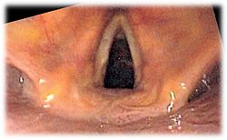
 تصویر آندوسکوپی از حنجره |
{{Infobox Anatomy
|Name         = حنجره
|Image        = Larynx external en.svg
|Caption      = کالبدشناسی حنجره، نمای قدامی-جنبی
|Image2       = Larynx endo 2.jpg
|Caption2     = تصویر [[آندوسکوپی]] از '''حنجره'''
}}
الگوهای وابسته
- {{جعبه اطلاعات استخوان}}
- {{جعبه اطلاعات ماهیچه}}
- {{جعبه اطلاعات رباط}}
- {{جعبه اطلاعات مغز}}
- {{Infobox Embryology}}
- {{Infobox neuron}}
- {{Infobox artery}}
- {{Infobox lymph}}
- {{Infobox Nerve}}
- {{Infobox vein}}


